Pelvic Ultrasound
What is a pelvic ultrasound?
A pelvic ultrasound is a non-invasive diagnostic exam that produces images that are used to assess organs and structures within the female pelvis. A pelvic ultrasound allows quick visualization of the female pelvic organs and structures including the uterus, cervix, vagina, fallopian tubes, and ovaries.
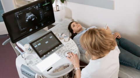
Ultrasound uses a transducer that sends out ultrasound waves at a frequency too high to be heard. The ultrasound transducer is placed on the skin, and the ultrasound waves move through the body to the organs and structures within. The sound waves bounce off the organs like an echo and return to the transducer. The transducer processes the reflected waves, which are then converted by a computer into an image of the organs or tissues being examined.
The sound waves travel at different speeds depending on the type of tissue encountered – fastest through bone tissue and slowest through the air. The speed at which the sound waves are returned to the transducer, as well as how much of the sound wave returns, is translated by the transducer as different types of tissue.
An ultrasound gel is placed on the transducer and the skin to allow for smooth movement of the transducer over the skin and to eliminate air between the skin and the transducer for the best sound conduction.
Another type of ultrasound is Doppler ultrasound, sometimes called a duplex study, used to show the speed and direction of blood flow in certain pelvic organs. Unlike a standard ultrasound, some sound waves during the Doppler exam are audible.
Pelvic ultrasound may be performed using one or both of 2 methods:
- Transabdominal (through the abdomen). A transducer is placed on the abdomen using the conductive gel
- Transvaginal (through the vagina). A long, thin transducer is covered with the conducting gel and a plastic/latex sheath and is inserted into the vagina
The type of ultrasound procedure performed depends on the reason for the ultrasound. Only one method may be used, or both methods may be needed to provide the information needed for diagnosis or treatment.
Other related procedures that may be used to evaluate problems of the pelvis include hysteroscopy, colposcopy, and laparoscopy.
.ashx?h=200&iar=0&mh=360&mw=520&w=200&hash=858D8DD88068FECC46AEA9F4BB0D4AB9)
What are female pelvic organs?
The organs and structures of the female pelvis are:
- Endometrium. The lining of the uterus
- Uterus (also known as the womb). The uterus is a hollow, pear-shaped organ located in a woman’s lower abdomen, between the bladder and the rectum. It sheds its lining each month during menstruation unless a fertilized egg (ovum) becomes implanted and pregnancy follows.
- Ovaries. Two female reproductive organs are located in the pelvis in which egg cells (ova) develop and are stored and where the female sex hormones estrogen and progesterone are produced.
- Cervix. The lower, narrow part of the uterus is located between the bladder and the rectum, forming a canal that opens into the vagina, which leads to the outside of the body.
- Vagina (also known as the birth canal). The passageway through which fluid passes out of the body during menstrual periods. The vagina connects the cervix and the vulva (the external genitalia).
- Vulva. The external portion of the female genital organs
What are the reasons for a pelvic ultrasound?
Pelvic ultrasound may be used for the measurement and evaluation of female pelvic organs. Ultrasound assessment of the pelvis may include, but is not limited to, the following:
- Size, shape, and position of the uterus and ovaries
- Thickness, echogenicity (darkness or lightness of the image related to the density of the tissue), and presence of fluids or masses in the endometrium, myometrium (uterine muscle tissue), fallopian tubes, or in or near the bladder
- Length and thickness of the cervix
- Changes in bladder shape
- Blood flow through pelvic organs
source: https://www.bothwellmedicalrooms.co.uk/